Osmotic Plasmolysis and the Wolfson Influence Explained
Understanding Plasmolysis: The Basics
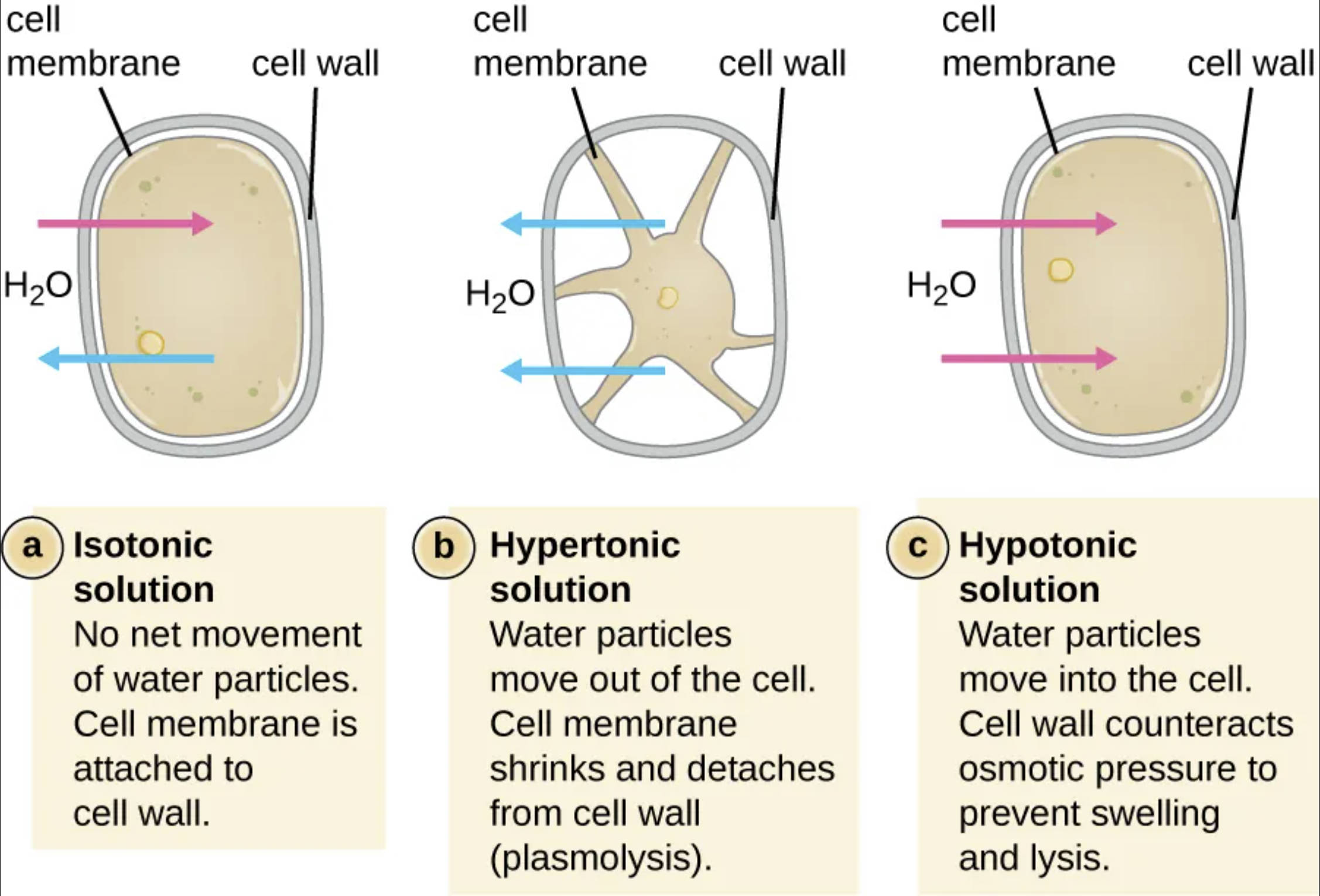
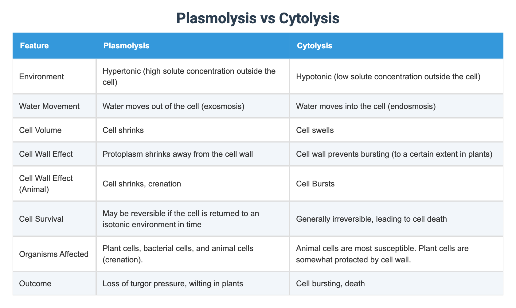
When a plant cell is placed in a hypertonic medium, water rushes out through osmosis, shrinking the cytoplasm and pulling the plasma membrane away from the rigid cell wall. The cell still retains some water, but its internal pressure—turgor—drops enough that the membrane detaches, a process botanists call plasmolysis. This phenomenon is a classic indicator of cellular water balance and a convenient tool for probing membrane integrity.
In the laboratory, plasmolysis is usually induced by adding a sucrose or mannitol solution to a leaf disc or protoplast preparation. The concentration of the external solution determines the rate and extent of water loss; higher osmotic potentials trigger faster and more pronounced membrane detachment. Researchers then monitor the changes using light microscopy, often staining the cell wall with dyes such as toluidine blue to keep the wall visible while the membrane recedes.
How Scientists Observe Plasmolysis
Microscopy remains the primary method for visualizing plasmolysis. A standard approach involves placing a thin tissue slice on a microscope slide, adding the osmotic solution, and recording the process under a high