Understanding Conus Medullaris Cysts: Causes, Symptoms, and Treatment Options
What Is a Conus Medullaris Cyst?
The conus medullaris is the tapered end of the spinal cord, typically located near the first or second lumbar vertebra. A cyst that forms in this region—sometimes called a perineural or syringomyelia‑type cyst—creates a fluid‑filled pocket that can press on nearby nerve fibers. Most people never notice such a cyst, but when it grows large enough to interfere with spinal pathways, pain, weakness, or sensory changes may appear.
Common Causes of Conus Medullaris Cysts
- Congenital malformations: Developmental anomalies, such as tethered cord syndrome, can leave a tiny space that fills with cerebrospinal fluid over time.
- Trauma: Even a mild spinal injury can disrupt the delicate lining of the central canal, prompting fluid accumulation.
- Inflammatory conditions: Diseases that inflame the meninges—like arachnoiditis—may block normal CSF flow and produce a cystic pocket.
- Degenerative spine disease: Osteophytes or disc herniations near the conus can create a one‑way valve effect, trapping fluid.
- Post‑surgical changes: Scar tissue after spinal surgery sometimes seals off a segment of the canal, leading to cyst formation.
In many cases, the exact trigger remains uncertain, and the cyst is discovered incidentally during imaging for an unrelated issue.
How Do These Cysts Present? Recognizing the Symptoms
Because the conus houses nerve roots that control the bladder, bowel, and lower limbs, symptoms tend to cluster around those functions. Typical complaints include:
- Sharp or burning pain that radiates to the buttocks, thighs, or calves.
- Gradual weakness in the legs, especially when walking uphill or climbing stairs.
- Altered sensation—numbness, tingling, or a “pins‑and‑needles” feeling—in the saddle region.
- Urinary urgency, frequency, or occasional incontinence.
- Constipation or a sense of incomplete evacuation.
Some patients experience only one of these signs, while others report a vague “tightness” in the lower back that worsens with activity. The variability often leads to misdiagnosis as sciatica or a simple disc problem.
Diagnosing a Conus Medullaris Cyst
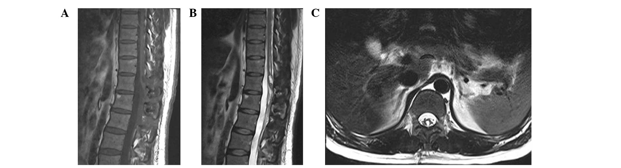
High‑resolution magnetic resonance imaging (MRI) remains the gold standard. On T2‑weighted scans, the cyst appears bright, mirroring cerebrospinal fluid, and its margins are well defined. Occasionally, a computed tomography (CT) myelogram is ordered to evaluate the exact communication between the cyst and the subarachnoid space.
Neurological examination helps localize the lesion: a positive Romberg sign, diminished ankle reflexes, or a “saddle anesthesia” pattern points directly to the conus region. When the diagnosis is unclear, a lumbar puncture may be performed to rule out infection or inflammatory disease.
Treatment Strategies: From Observation to Surgery
Conservative management is the first line for small, asymptomatic cysts. Regular MRI follow‑ups every six to twelve months track any growth, while physical therapy focuses on core stability and gentle stretching to reduce pressure on the lower spine.
When pain, weakness, or bladder dysfunction begins to interfere with daily life, interventional options are considered. The most common procedure is a cyst fenestration or drainage performed through a minimally invasive posterior approach. Surgeons create a small opening in the cyst wall, allowing fluid to re‑enter the normal CSF pathway. In many cases, a tiny shunt is placed to prevent re‑accumulation.
For cysts that are tightly adhered to the conus or that recur after drainage, complete excision may be necessary. This carries a higher risk of nerve injury, so it is reserved for patients with progressive neurological decline despite previous attempts.
Post‑operative care emphasizes gradual mobilization, bladder training, and routine imaging to confirm that the cyst remains collapsed. Most people report significant symptom relief within weeks, though a minority experience lingering paresthesias that gradually improve over months.
Living With a Conus Medullaris Cyst: Practical Tips
Even after successful treatment, lifestyle adjustments can help prevent flare‑ups. Maintaining a healthy weight reduces stress on the lumbar spine, while low‑impact aerobic activities—such as swimming or stationary cycling—keep the surrounding musculature strong without jarring the delicate nerve roots.
Staying hydrated supports normal cerebrospinal fluid dynamics, and avoiding prolonged sitting or heavy lifting can minimize pressure spikes. If bladder symptoms persist, a timed voiding schedule and pelvic floor exercises often make a noticeable difference.
Lastly, keep an eye on warning signs: sudden worsening of leg strength, new onset incontinence, or a rapid increase in back pain should prompt an immediate call to your physician. Early intervention can forestall permanent nerve damage.
Frequently Asked Questions
- Can a conus medullaris cyst disappear without treatment? Rarely. Small cysts sometimes shrink as CSF flow normalizes, but most remain stable or grow slowly, so periodic imaging is advised.
- Is surgery always required? No. Many patients are managed conservatively, especially if the cyst is tiny and asymptomatic. Surgery is reserved for those with progressive neurological deficits or severe pain.
- What is the success rate of cyst drainage? Studies suggest that about 70‑80% of patients experience meaningful symptom relief after a well‑performed fenestration, though a small percentage may need repeat procedures.
- Will I need lifelong follow‑up? Ongoing monitoring is recommended, typically with MRI every 1‑2 years, to catch any recurrence early.